

On June 2, 2020, a paper entitled “Medial prefrontal cortex represents the object-based cognitive map when remembering an egocentric target location” was online published in Cerebral Cortex by Dr. Yuji Naya group in the School of Psychological and Cognitive Sciences at Peking University and the PKU-IDG/McGovern Institute for Brain Research.
During navigation, it is necessary to locate our self-position in the current spatial environment as well as to locate the objects relative to the self-body (i.e., egocentric location). To conduct each of the two mental operations, we need map-like representations, called “cognitive map” (Tolman 1948). After the discovery of “place cells,” the hippocampus (HPC) of the medial temporal lobe (MTL) has been considered responsible for the cognitive map, and crucial contributions of the HPC to spatial memory have also been reported by animal model as well as human studies. However, it remains largely unknown how neural substrates of the cognitive map are involved in the two mental operations required to locate specific objects within the environment. Despite extensive studies on the spatial elements related to the cognitive map (e.g., self-location, head-direction etc.) (O'Keefe and Dostrovsky 1971; Vass and Epstein 2013), there is still a lack of sufficient isolation and characterization of the neural signal of the cognitive map under the previous research paradigms.
To address these issues, we devised a novel 3D spatial-memory task with spatial environments defined by three different human characters, which would enable us to identify the representation of the cognitive map and to investigate how it is related to the two mental operations (Fig. 1a). The spatial arrangement of the characters was referred to as the “map” (Fig. 1b). Using representational similarity analysis (RSA), we investigated the brain regions that code the maps when the participants located themselves in the virtual environment (facing period) and when they recalled an egocentric location of a target character (targeting period). We found the neural representation of object-based maps in the hippocampus (HPC) during the facing period, while the medial prefrontal cortex (mPFC) represented it during the targeting period (Fig. 2). These results provides the first experimental evidence demonstrating the mental representations of the coherent space formed by mobile objects (e.g., humans) during the spatial navigation. The contribution of the object-based cognitive map may generalize to other aspects of cognition, such as navigating social interactions.
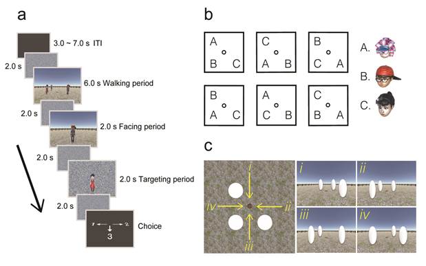
Fig. 1 (a) Spatial-memory task paradigm. Each trial consisted of three periods. In the walking period, participants walked toward three human characters using the first-person perspective and stopped on a wood plate. In the facing period, one of the human characters was presented, indicating the participant’s current self-orientation. In the targeting period, a photo of another character was presented. Participants chose the direction of the target character relative to their body upon presentation of a response cue. (b) Maps were defined by the relative position of the three human characters. (c) The walking directions were defined by the spatial layout of the three human characters from the participant’s first-person perspective.

Fig. 2 The neural representation of cognitive map during the facing and targeting period.
Article link: https://doi.org/10.1093/cercor/bhaa117
This study was supported by National Natural Scientific Foundation of China (NSFC). The corresponding author is Dr. Yuji Naya. The first author is Bo Zhang, a Ph.D. student from the School of Psychological and Cognitive Sciences at Peking University.
Reference
Tolman, E. C. (1948). Cognitive maps in man and animals. Psychol Rev, 55, 189-208.
O'Keefe, J., & Dostrovsky, J. (1971). The hippocampus as a spatial map: preliminary evidence from unit activity in the freely-moving rat. Brain research.
Vass, L. K., & Epstein, R. A. (2013). Abstract representations of location and facing direction in the human brain. Journal of Neuroscience, 33(14), 6133-6142.